Field-Cycling Imaging (FCI): A Game-Changer for the Early Detection of Osteoarthritis
23 January 2025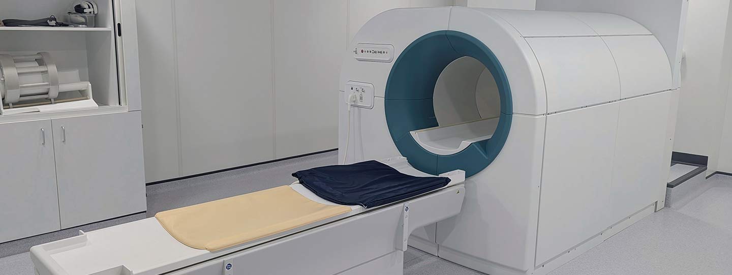
A new imaging technique called Field-Cycling Imaging (FCI) offers hope for earlier detection and improved monitoring of osteoarthritis, potentially transforming how we understand and treat this condition.
Why is the early detection of osteoarthritis so important?
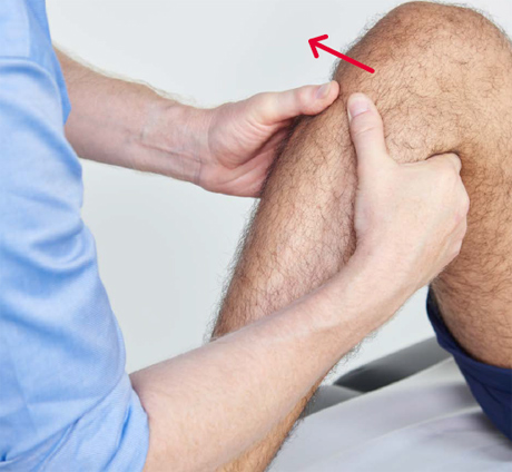
Usually, the early signs of knee osteoarthritis (OA) go undetected until significant joint damage has occurred. Current detection methods are not sensitive enough to identify the condition in its early stages and we lack clear indicators of its onset.
The early detection of knee OA is critical to slowing the disease's progression and improving people’s quality of life. Currently, there are no diagnostic methods that can reliably detect OA in its early stages, which means many people miss the opportunity for timely intervention.
The inability to detect changes in the knee at an early stage is partly the reason why there are still no treatments which can slow down the progression of OA. Being able to detect OA and monitor its progress may open the door to new treatment options before the disease progresses further.
A breakthrough in early detection
Previously, researchers at the University of Aberdeen discovered that a property of the cartilage of the knee which determines response to different magnetic fields, is starkly different in osteoarthritic cartilage compared to healthy cartilage.
This finding could mark a key breakthrough in early detection. Identifying changes in how the cartilage responds to different magnetic fields before damage occurs could be a crucial indicator of OA onset.
This discovery was made possible through a new technology called Field-Cycling Imaging (FCI).
What is Field-Cycling Imaging (FCI) and how does it work?
Developed at the University of Aberdeen, FCI follows in the footsteps of MRI (Magnetic resonance imaging). The first MRI scanner was also invented at the University of Aberdeen around 50 years ago and has since gone on to save millions of lives.
FCI works similarly to MRI but with one key difference: FCI can very quickly change the strength of the magnetic field during the scan. Traditional MRI uses a constant magnetic field throughout the body to create images, while FCI can adjust the magnetic field at different points during the scan.
Dr James Ross from the University of Aberdeen explains:
“Being able to change the strength of the magnet allows us to see how different tissues respond to different magnetic fields. "
"We already know from our research that the way that tissues respond to different magnetic fields changes in disease, potentially at quite an early stage. Because these changes are sensitive to disease, this information might potentially be used as a new diagnostic 'biomarker' for many conditions, including osteoarthritis.”
The MRI scanners you might find in a hospital cannot change their magnetic fields which means that this information is completely invisible to them.
Detecting changes in tissue response to different magnetic fields is especially important for conditions like knee OA, where early detection can lead to better treatment outcomes.
“It is exciting to be part of the team who brought MRI to the world and now with FCI technology, the possibilities are endless for disease detection and diagnosis”
PioKneering the future of knee osteoarthritis detection
Versus Arthritis has funded the PIOKNEER study, led by Professor Cosimo de Bari, who recently moved from the University of Aberdeen to the University of Edinburgh, to further investigate the potential of FCI to detect early knee OA. Previously, researchers had studied small samples of cartilage from people undergoing joint replacement surgery. With the PIOKNEER study they will move from these cartilage samples to scan living people.
The researchers will track individuals who are experiencing knee pain but who have no visible signs of OA. Through annual FCI scans, researchers will look for early signs of disease progression, identifying the moments when FCI might be most useful for early diagnosis.
What will the PIOKNEER study investigate?
- Monitor Disease Progression: Track knee OA over time using both X-rays and FCI to understand when changes first appear. Researchers will also collect samples from study participants to measure indicators of joint damage (biomarkers).
- Identify Early Indicators: Pinpoint how cartilage responds to different magnetic fields at the earliest stages of knee OA.
- Develop Effective Treatments: Help determine which treatments are most effective by linking disease progression with treatment response. Selected participants will be invited to undergo small knee biopsies for tissue analyses in the laboratory to better understand the disease at an early stage and help identify new opportunities for targeted therapeutic intervention.
- Improve care: The PIOKNEER project will prioritise the patient experience, by exploring the gaps in care pathways in close collaboration with people with lived experience.
FCI: complementing, not replacing, existing technologies
While FCI is a powerful new tool, it is not designed to replace traditional MRI or other imaging techniques. Instead, it complements existing methods by providing additional, unique insights that current technologies cannot capture.
Just as ultrasound and X-rays offer different perspectives on a patient's health, FCI will add another layer of valuable information to help doctors understand knee OA in more detail.
So, what’s next?
The PIOKNEER study marks the beginning of a new era in our understanding of knee OA diagnosis and treatment. As researchers continue to study the effectiveness of FCI, this innovative technology has the potential to become a standard tool in clinical practice. The results from this study will be compared with similar research worldwide, ensuring that the best possible approaches to the early detection of OA are developed.
Do you want to stay updated on the latest advancements in knee osteoarthritis research?
If you would like to be informed about the latest information regarding Versus Arthritis research you can sign up for updates or you can explore our research on our website. You can also support these life-changing studies by making a donation.
Donate to the ongoing research supported by Versus Arthritis